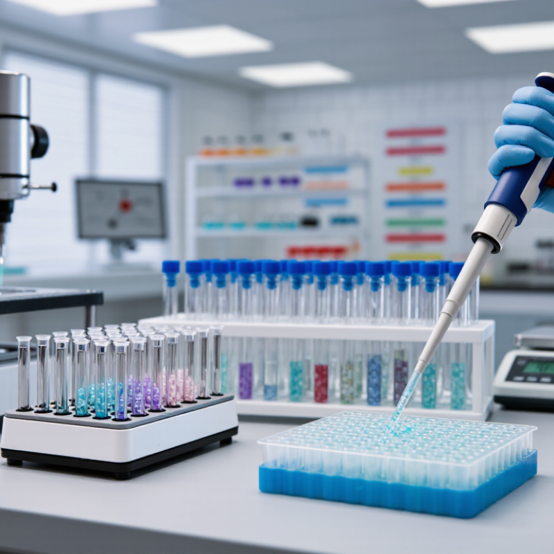
Choosing the 2026 best magnetic beads for size selection requires more than comparing recovery percentages. Magnetic Beads Size Selection influences fragment distribution, adapter removal, sequencing yield, and downstream data quality. In a routine library-preparation workflow, a small ratio error can leave unwanted 150 bp fragments beside the intended 350 bp library. That difference matters.
Market reports support the growing importance of this workflow. MarketsandMarkets’ Magnetic Beads Market—Global Forecast to 2029 identifies expanding demand from genomics, diagnostics, and automated sample preparation. Grand View Research’s nucleic acid isolation and purification analysis also highlights automation and reproducible purification as major market drivers. However, these reports measure broad market activity, not the performance of every bead formulation. Their forecasts can differ.
That limitation deserves attention.
A useful 2026 comparison should examine binding chemistry, size-selection sharpness, recovery, lot consistency, liquid-handling compatibility, and total cost per sample. Practical evidence matters. A bead system may perform well with short PCR products but show weaker separation when input DNA is scarce or highly fragmented. Experienced laboratory teams also check pipetting dead volume, magnetic stand geometry, ethanol carryover, and drying time. These details often decide whether a protocol works on Monday morning.
This guide will compare leading options through an evidence-based and application-focused lens. It will consider published specifications, technical documentation, independent workflow data, and realistic laboratory constraints. No single product is best for every library. The strongest choice is the one that delivers predictable fragment selection within your sample type, instrument setup, and validation requirements.
Magnetic beads make DNA and RNA size selection practical by turning fragment length into a controllable binding event. Polyethylene glycol and salt help nucleic acids attach to bead surfaces. Larger fragments usually bind at lower bead ratios, while smaller fragments remain in solution. This principle supports library cleanup, adapter removal, and targeted fragment recovery.
According to Grand View Research’s 2024 Magnetic Beads Market report, the sector was estimated near USD 2.7 billion in 2023, with projected annual growth above 11% through 2030. MarketsandMarkets also forecasts strong expansion for nucleic acid isolation technologies. The demand reflects a real laboratory need: repeatable cleanup with less centrifugation and smaller sample losses.
In practice, a 0.6× bead ratio may retain larger DNA while leaving shorter fragments in the supernatant. The exact cutoff changes with salt, PEG, fragment concentration, and incubation time. RNA needs extra care because it degrades easily and often contains highly similar fragment sizes. Use low-binding tubes, short handling times, and validated ratios. Small mistakes matter.
A clean tube is not proof of clean selection. Measure fragment distribution after cleanup. Fluorescence alone cannot show whether short adapters remain. This is where many protocols feel reliable, yet still need review. Calibration with a size ladder and recovery controls gives stronger evidence than habit.
Magnetic-bead size selection uses polyethylene glycol and salt to reversibly bind nucleic acids to paramagnetic particles. Adjusting the bead-to-sample ratio changes the approximate fragment-size threshold, allowing smaller or larger DNA and RNA molecules to be retained or removed. The chart shows representative size targets commonly handled in bead-based workflows; actual recovery depends on bead chemistry, buffer composition, nucleic-acid concentration, and protocol conditions.
2026 Best Magnetic Beads for Size Selection?
Choosing magnetic beads in 2026 requires more than comparing price or particle size. The bead-to-sample ratio controls the fragment cutoff, while salt and polyethylene glycol levels influence DNA binding. A 2024 magnetic-beads market outlook reports expected annual growth above 10% through 2030, reflecting wider use in sequencing and automated workflows. Demand is rising. Consistency matters more.
Experienced laboratories should examine recovery, size-selection accuracy, carryover, and lot-to-lot variation. Check the manufacturer’s fragment-size curves, not only the headline specification. A 0.8x ratio may remove smaller fragments, but it is not universal. Different DNA inputs can shift the cutoff. That detail is easy to miss. High-quality beads should produce clean separation with low residual liquid after washing. Compatibility with robotic magnets also matters, especially when volumes fall below 20 microliters. The 2024 NGS Workflow Survey reported that workflow standardization remains a major laboratory priority, supporting stronger bead validation before routine use.
Tips: Test three ratios around the target cutoff. Use a defined DNA ladder and measure recovery with fluorometry. Confirm fragment profiles by electrophoresis when precision matters. Record mixing time, drying time, and elution temperature. Small timing differences can change results. I would also challenge the assumption that the highest recovery is best; oversized fragments or adapter dimers may still remain. Review performance after several lots, not one attractive pilot run.
2026 Best Magnetic Beads for Size Selection?
Comparing Bead Types for Different Fragment Size Ranges
The best magnetic beads depend on the fragment window, not a single “universal” product. Standard carboxylated beads work well for routine cleanup and broad DNA selection. At about 0.8–1.8× bead-to-sample ratios, they can retain many fragments while removing primers, salts, and short by-products. Higher ratios usually capture smaller fragments. Lower ratios favor larger DNA.
For narrow windows, dual-sided selection is more practical. A low-ratio binding step removes very large fragments, while a second, higher-ratio step captures the desired middle range. Some high-retention bead formulations improve recovery of long DNA, but they may also increase viscosity and transfer contaminants. Handle them gently. Large fragments can shear during repeated pipetting.
I check the result with a fragment analyzer or gel, especially when the library contains many short peaks. The manufacturer’s ratio is only a starting point; buffer composition, DNA concentration, and mixing time change the cutoff. In my experience, a 0.1× adjustment can noticeably shift recovery. That detail is easy to overlook. For fragments below 200 bp, use a bead type validated for short-fragment discrimination and extend magnetic separation until the liquid clears. I still run a small pilot first, because the cleanest theoretical size range can perform poorly with unusually GC-rich or low-input samples.
Magnetic bead size selection begins with the target fragment range, not a favorite bead product. In my bench experience, clean the sample before changing bead ratios. Remove visible debris and confirm concentration with a suitable fluorometric method. Record sample volume, buffer composition, and expected fragment sizes. Small details matter here. A ratio that works for one library may fail in another.
Use a calibrated pipette to mix the sample with beads at a measured ratio. Lower ratios often retain larger fragments, while higher ratios recover smaller fragments. Treat this as a starting point, not a guarantee. Mix gently for 10–15 minutes at room temperature, avoiding foam. Place the tube on a magnet until the liquid clears. Transfer the supernatant or retain the pellet, depending on the desired size fraction. Label both fractions; discarded liquid can reveal unexpected loss.
Wash the magnetic pellet twice with freshly prepared ethanol-based wash solution while keeping it on the magnet. Do not disturb the pellet. Remove residual wash carefully, then air-dry briefly. Overdrying can make elution stubborn. Elute in low-salt buffer or water, mix gently, and incubate for several minutes. Measure recovery and inspect fragment distribution with an appropriate analytical method. If the profile is too broad, adjust the ratio in small increments. I sometimes change too many variables at once; that weakens comparison. Keep one control sample and document every pause, temperature, and transfer step.
| Category | Workflow Step or Parameter | Recommended Setting | Typical Value or Size Range | Purpose and Expected Result | Key Handling and Quality-Control Notes |
|---|---|---|---|---|---|
| 1. Magnetic Bead Selection Criteria | |||||
| Bead chemistry | Particle surface | Use carboxylated paramagnetic particles compatible with PEG and salt-mediated nucleic-acid binding. | Carboxyl-functionalized magnetic particles | Enables reversible DNA binding during the binding and washing steps. | Confirm that the bead formulation is intended for nucleic-acid purification or size selection rather than protein purification. |
| Particle size | Magnetic particle diameter | Use a uniform particle suspension with efficient magnetic capture. | Commonly about 0.5–1.0 µm for SPRI-type workflows | Provides a balance between binding surface area, suspension stability, and rapid magnetic separation. | Particle diameter alone does not determine the DNA fragment cutoff; buffer chemistry and bead-to-sample ratio are more influential. |
| Bead suspension | Resuspension before use | Mix thoroughly until the suspension is visibly homogeneous. | Invert, vortex briefly, or pipette-mix according to the bead instructions | Ensures consistent bead concentration and reproducible size selection. | Avoid creating excessive foam. Beads settle quickly, so remix immediately before dispensing each aliquot. |
| Buffer compatibility | Binding environment | Use a PEG/salt-based binding buffer or a validated equivalent. | PEG and salt concentrations are chemistry-dependent | Controls the minimum DNA length that can bind to the beads. | Do not assume that a ratio calibrated for one formulation will produce the same cutoff with another formulation. |
| 2. Step-by-Step Magnetic Bead Size Selection Workflow | |||||
| Sample preparation | Normalize the DNA sample | Measure sample volume and, when possible, DNA concentration before adding beads. | Use a consistent input volume for comparable reactions | Improves reproducibility because DNA concentration can affect binding behavior and recovery. | Remove detergents, high EDTA levels, residual ethanol, and other contaminants that may interfere with binding or downstream enzymatic reactions. |
| Ratio selection | Choose the bead-to-sample ratio | Calculate the bead volume as a multiple of the DNA sample volume. | 0.6×–1.8× commonly used for DNA size selection | Lower ratios preferentially retain larger fragments; higher ratios retain progressively smaller fragments. | Use a validated ratio curve for the specific bead chemistry and library type. |
| Large-fragment enrichment | Low-ratio selection | Add beads at a low ratio, mix thoroughly, and retain the bead-bound fraction. | 0.6×; often excludes fragments below approximately 250–400 bp | Useful when removing short adapter dimers or very small degradation products from a DNA library. | The approximate cutoff varies substantially with DNA mass, salt concentration, and the bead formulation. |
| Moderate size selection | Intermediate-ratio selection | Use a moderate bead ratio when removing smaller fragments while retaining most desired library molecules. | 0.8×; often excludes fragments below approximately 150–250 bp | Commonly used for short-read sequencing library cleanup and removal of small by-products. | Verify the final distribution with electrophoresis or a fragment analyzer when fragment size is critical. |
| Broad DNA recovery | Near-total-binding selection | Use a higher ratio to recover a broad range of DNA fragments. | 1.0×; often retains fragments above approximately 100–150 bp | Provides a general-purpose cleanup with relatively high recovery of typical library fragments. | May retain adapter dimers and other short products, depending on the chemistry and sample composition. |
| Maximum small-fragment retention | High-ratio selection | Use a high ratio when the protocol requires retention of shorter DNA fragments. | 1.2×–1.8×; commonly retains fragments below the cutoff of lower-ratio conditions | Useful for broad-range DNA recovery or workflows where small fragments are part of the desired product. | Higher ratios increase reagent use and may also increase carryover of short contaminants. |
| Mixing | Bead–sample binding | Mix by pipetting until the sample is uniform and no visible bead clumps remain. | Approximately 10–15 mixing cycles or brief controlled vortexing | Promotes even contact between DNA molecules and bead surfaces. | Use the same mixing method and intensity across samples to reduce variability. |
| Binding incubation | Allow DNA to bind | Incubate the mixed suspension before magnetic separation. | Typically 5–10 minutes at room temperature | Allows sufficient time for DNA adsorption under the selected PEG/salt conditions. | Longer incubation does not always improve recovery and can increase nonspecific carryover in some samples. |
| Magnetic separation | Capture the beads | Place the tube or plate on a suitable magnetic rack and wait until the liquid becomes clear. | Typically 2–5 minutes | Collects the DNA-bound beads against the tube wall or well edge. | Do not aspirate until the bead pellet or bead layer is stable and clearly separated from the liquid. |
| Supernatant handling | Discard or retain the liquid fraction | Determine in advance whether the target DNA is in the bead-bound fraction or the discarded supernatant. | Bead-bound fraction for standard cleanup; supernatant for some double-sided selections | Prevents accidental loss of the desired size range. | For double-sided selection, the first low-ratio supernatant may contain the smaller fragments that are subsequently captured with a higher bead ratio. |
| First wash | Wash with fresh ethanol-based wash solution | Wash the bead pellet without disturbing it. | Usually 70–80% ethanol | Removes PEG, salts, and soluble contaminants while DNA remains bound to the beads. | Use freshly prepared ethanol wash solution and avoid prolonged exposure that can make the pellet difficult to resuspend. |
| Second wash | Repeat ethanol wash | Perform a second wash using the same ethanol concentration. | One additional wash is commonly used | Improves removal of residual salts and PEG. | Keep the bead pellet immobilized during aspiration to minimize sample loss. |
| Drying | Remove residual ethanol | Air-dry only until visible liquid ethanol has evaporated. | Typically 2–5 minutes, depending on pellet size and vessel format | Reduces ethanol carryover into the elution and downstream reaction. | Over-drying can make beads difficult to resuspend and may reduce DNA recovery. |
| Elution | Release purified DNA | Add a low-salt elution buffer or nuclease-free water and resuspend completely. | Common elution volume: 15–50 µL | Recovers the size-selected DNA for amplification, library preparation, or analysis. | Use a smaller volume for concentration and a larger volume when maximum recovery is more important. |
| Elution incubation | Allow DNA to enter the liquid phase | Incubate the resuspended beads before magnetic separation. | Typically 2–5 minutes at room temperature | Improves release of DNA from the bead surface. | Mix gently during incubation if the protocol permits, then separate the beads magnetically before transferring the eluate. |
| Final recovery | Transfer the eluate | Move the clear eluate to a clean tube without carrying over beads. | Recover the full usable eluate volume | Produces the final size-selected DNA sample. | Residual beads, ethanol, PEG, or salt can inhibit polymerases and distort downstream quantification. |
| 3. Selection Verification and Troubleshooting | |||||
| Fragment-size verification | Assess the final DNA distribution | Use an appropriate electrophoretic or microfluidic fragment-analysis method. | Compare observed distribution with the expected cutoff range | Confirms whether the selected ratio achieved the intended size profile. | Use the same analysis method and ladder range when comparing different conditions. |
| Low recovery | Investigate poor yield | Check bead resuspension, binding time, ethanol carryover, over-drying, and elution mixing. | Recovery is chemistry- and input-dependent | Identifies procedural causes of DNA loss rather than assuming the ratio is incorrect. | Include a known-control sample when optimizing a new bead formulation or sample type. |
| Too many short fragments | Tighten the lower-size cutoff | Reduce the bead ratio incrementally or use a validated double-sided selection. | Test neighboring ratios, such as 0.6×, 0.7×, and 0.8× | Removes more short fragments while retaining the desired larger molecules. | Optimize empirically because a small ratio change can produce different results across bead chemistries. |
| Loss of desired small fragments | Increase the lower-size retention | Increase the bead ratio incrementally and confirm the resulting profile. | Test neighboring ratios, such as 1.0×, 1.2×, and 1.5× | Improves recovery of shorter target fragments. | Higher ratios may also retain adapter dimers and other low-molecular-weight contaminants. |

The best magnetic beads for size selection are not defined by recovery alone. They must produce a narrow fragment window, stable yield, and clean downstream reactions. In practice, bead-to-sample ratio controls the cutoff more strongly than many users expect. A small pipetting error can shift recovery by dozens of base pairs.
Common errors include incomplete mixing, over-drying, and disturbing the pellet during ethanol removal. Dry beads can become difficult to resuspend. That often creates uneven library concentrations. The 2024 Association of Biomolecular Resource Facilities Core Facilities Survey highlights growing demand for standardized sequencing workflows and stronger documentation. That finding matches routine laboratory experience: undocumented timing causes avoidable variation. I still see teams changing incubation times without recording room temperature.
Use calibrated pipettes and mix until the suspension looks uniform. Keep binding time consistent, usually around five to ten minutes. Avoid aspirating the bead surface. For narrow size selection, test three ratios across a small pilot plate instead of trusting one published setting. It costs less than repeating a full library preparation.
Quality control should combine fluorometric concentration, electrophoretic size profiles, and fragment-size distribution. Include a no-template or blank process control when contamination is plausible. The Genome in a Bottle benchmarking reports emphasize coverage, insert-size consistency, and library complexity as meaningful sequencing checks. A practical rule is to repeat a batch when replicate concentrations vary beyond 10 percent or when the modal fragment size shifts unexpectedly. These thresholds are useful, not sacred. Some laboratories accept wider variation, and that may be reasonable for low-complexity samples.